Learning Objective
By the end of this lesson, you will be able to identify the heart’s normal pacemaker hierarchy, state the expected intrinsic rate range of the SA node, AV junction, and ventricular escape focus, and explain what it means when a lower backup pacemaker takes over.
Opening Hook
You are watching the monitor when a patient’s rate slows from the 70s into the 30s, but the strip does not go flat. Something is still firing. That moment only makes sense if you know the heart has a built-in pacing hierarchy instead of a single all-or-nothing starter switch. This lesson shows you who normally leads, who backs up the system, and why a slow rhythm can still be a lifesaving escape rhythm rather than electrical silence.
What To Notice First
Notice the hierarchy before the numbers. The SA node normally wins because it fires fastest. The AV junction can take over if the SA node slows too much or its impulses do not reach the ventricles. A ventricular escape focus sits even lower and usually fires slowest, but it can still keep the patient from immediate asystole.
The Heart Does Not Have Only One Possible Pacemaker
Earlier in this module, you learned the normal route of conduction and how that route becomes a visible waveform. Now add one more rule: key parts of the heart’s conduction system have automaticity, which means they can depolarize on their own without waiting for an outside nerve to trigger them.
That does not mean every area should lead all the time. In a healthy resting heart, the sinoatrial node, or SA node, acts as the dominant pacemaker because it normally depolarizes faster than the backup sites below it. Faster tissue suppresses slower tissue. As long as the SA node is firing reliably and the impulse conducts normally, the lower pacemakers stay quiet.
The SA Node Is the Normal Leader
The SA node sits high in the right atrium near the entry of the superior vena cava. In a resting adult, its intrinsic firing rate is usually about 60 to 100 beats per minute. When you later identify normal sinus rhythm, sinus bradycardia, or sinus tachycardia, you are still talking about rhythms that begin in this same location.
What makes a beat sinus is not only the rate. It is the point of origin. A sinus beat starts in the SA node, spreads through the atria, reaches the AV node, pauses briefly there, and then continues through the His-Purkinje system into the ventricles.
That is why sinus rhythm usually gives you the familiar sequence you already know: a P wave before each expected QRS complex, with the atria leading the ventricles in an organized way.
The AV Node and AV Junction Are the Usual Backup
The atrioventricular node, or AV node, normally serves as the gatekeeper between atria and ventricles. It slows conduction briefly so the ventricles have time to fill. But it is not only a relay station. Tissue in and around the AV junction can also act as a backup pacemaker.
Its intrinsic rate is usually around 40 to 60 beats per minute. That is slower than the SA node, which is why it normally stays in the background. If the SA node fails to fire, fires too slowly, or its impulses do not get through, the AV junction may escape and take control.
At this stage, the main concept is hierarchy, not detailed rhythm recognition. Later you will learn how junctional rhythms can change P-wave appearance, hide P waves, or even produce retrograde atrial activation. For now, the key point is simpler: if a backup pacemaker lower than the SA node takes over, the rhythm often slows because the backup site’s natural rate is slower.
Ventricular Escape Is the Last Built-In Safety Net
Below the AV junction, distal conduction tissue and the ventricles can also generate impulses. This is the slowest built-in backup, often around 20 to 40 beats per minute. When that lower focus starts the beat, it is called a ventricular escape rhythm.
This matters clinically because a ventricular escape rhythm is usually not the heart’s first choice. It is the heart’s emergency fallback when higher pacemakers are not leading effectively. A slow ventricular escape rhythm may be enough to preserve some output for a short time, but it is also a sign that the normal pacing hierarchy above it has failed or been blocked.
That is why the phrase escape rhythm is so important. It means the lower pacemaker is not merely being irritable or premature. It is rescuing the rhythm after a pause or failure from above.
Rate Is the Clue That Reveals the Hierarchy
One practical way to remember the hierarchy is to connect each pacemaker level to its typical resting rate range:
- SA node: typically about 60 to 100 beats per minute
- AV junction: typically about 40 to 60 beats per minute
- Ventricular escape focus: typically about 20 to 40 beats per minute
These ranges are not magic labels that diagnose every strip by themselves. They are orientation tools. If a rhythm is unexpectedly slow, you should start thinking about whether a lower pacemaker has taken over. If the rhythm is very slow and the QRS later turns out to be wide, ventricular origin becomes even more plausible, but the full recognition rules belong to later modules.
At this point in the course, the best habit is to ask one simple question: Who is pacing this heart right now?
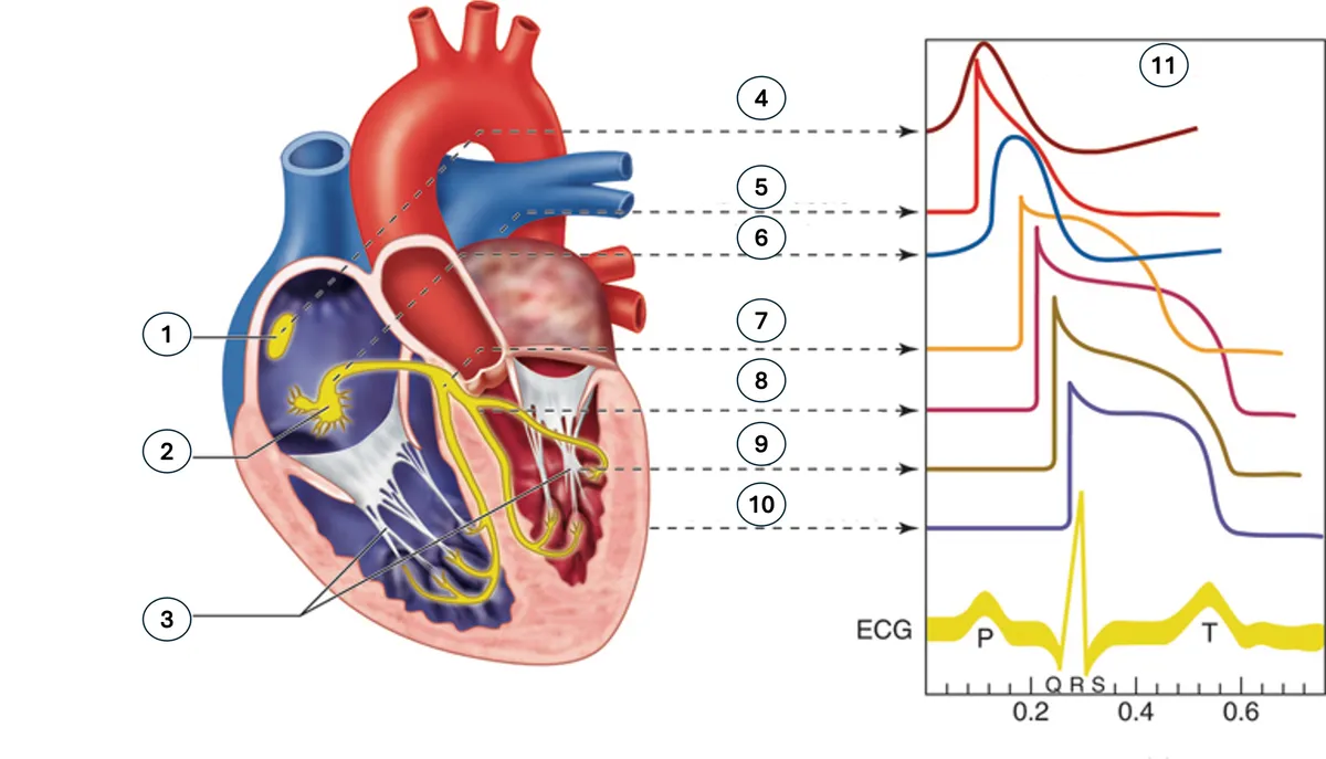
Open image key
Pacemaker Hierarchy and Conduction System
- 1
- Sinoatrial node (SA node)
- 2
- Atrioventricular node (AV node)
- 3
- Purkinje system
- 4
- SA node action potential
- 5
- Atrial muscle
- 6
- AV node
- 7
- Common bundle (bundle of His)
- 8
- Bundle branches
- 9
- Purkinje fibers
- 10
- Ventricular muscle
- 11
- Action potential inset
This conduction map helps you visualize why the higher sites usually lead and why lower escape sites produce backup rhythms only when normal control from above is lost.
Worked Example: Who Took Over After the Pause?
Imagine you are watching a strip that had been marching along normally. Then there is a pause. After that pause, a beat finally appears, but it is much later than expected.
Your first question is not the exact diagnosis yet. Your first question is where the rescue beat probably came from in the hierarchy.
If the next active pacemaker is the AV junction, you would expect a backup rhythm that is slower than normal sinus but not usually as slow as a ventricular escape focus. If the next active pacemaker is in the ventricles, you would expect an even slower fallback rate.
The teaching point is not to memorize a single strip pattern here. It is to see the logic. Higher pacemaker fails. Lower pacemaker waits out its own intrinsic cycle length. Then it fires to keep the heart from remaining electrically silent.
That is the meaning of an escape rhythm in one sentence: a slower backup pacemaker fires because the faster normal pacemaker did not lead the next beat.
Why Lower Pacemakers Usually Stay Quiet Until They Are Needed
The reason backup pacemakers do not usually compete on every beat is called overdrive suppression. When the SA node keeps sending impulses at a faster rate, the slower pacemakers below it are reset before they can finish their own cycle and fire.
When that faster input disappears or fails to conduct, the suppressed pacemaker below is finally able to depolarize on its own. That is why backup pacing appears after a failure above, not during stable normal sinus rhythm.
You do not need ion-channel detail for this lesson. You do need the concept, because it explains why the pacing hierarchy is orderly instead of chaotic most of the time.
What This Means at the Bedside
At a monitoring level, the pacing hierarchy helps you interpret urgency without overcalling every slow rhythm the same way.
- A rhythm from the SA node may still be slow, but the normal leader is intact.
- A rhythm from the AV junction means the normal leader above is not currently in charge.
- A ventricular escape rhythm means the heart is relying on its last built-in backup.
The patient may still have a pulse with any of these, but a lower escape site usually raises more concern that the normal conduction system above has failed or that conduction to the ventricles is seriously impaired. You always match the strip to perfusion, symptoms, blood pressure, and the overall clinical picture.
Don’t Confuse This With
- Sinus bradycardia: Both can be slow rhythms. The best distinguishing clue is origin: sinus bradycardia still starts in the SA node, while an escape rhythm starts from a lower backup pacemaker.
- A premature beat: Both involve a site outside the usual timing of the normal rhythm. The best distinguishing clue is timing: a premature beat comes too early, while an escape beat comes late after a pause or failed impulse from above.
- Asystole: Both can appear during dangerous bradycardic situations. The best distinguishing clue is that an escape rhythm still shows organized electrical activity from a backup pacemaker, while asystole means there is no meaningful cardiac electrical activity to record.
Lesson Summary
- The heart has a pacing hierarchy, not a single all-or-nothing trigger site, because multiple cardiac tissues have automaticity.
- The SA node is normally the dominant pacemaker because it usually fires fastest at about 60 to 100 beats per minute.
- The AV junction is the usual backup pacemaker and typically fires at about 40 to 60 beats per minute when the SA node fails or its impulses do not conduct normally.
- Ventricular escape is the slowest built-in backup, usually around 20 to 40 beats per minute, and appears when higher pacemakers are not effectively controlling the rhythm.
- Escape rhythms are protective because they prevent prolonged electrical silence, but they also signal that the normal pacing system above is not leading as expected.
- A slow rhythm is not enough to label the pacemaker source by itself, but the rate range gives you a strong first clue about where in the hierarchy the impulse may be coming from.
What’s Next
The next lesson explains why the same electrical event can look different depending on which lead records it. Once you know who started the beat, you are ready to see why that beat’s shape still changes from one viewing angle to another.