Learning Objective
By the end of this lesson, you will be able to trace normal blood flow through the heart and describe how the electrical impulse travels from the sinoatrial node to the ventricles.
Opening Hook
You are looking at a monitor and someone asks a simple question: did that beat start in the atria and travel normally, or did it start lower in the heart? You cannot answer that from waveform shape alone if the heart is still a blur of chambers, valves, and arrows. This lesson gives you the map first. Once you know where blood goes and where the electrical signal normally travels, the ECG stops looking like abstract spikes and starts looking like a real event happening in a real organ.
The Heart Is Two Pumps Working in Sequence
Your heart is a muscular four-chamber pump with a right side and a left side. The atria are the two upper chambers that receive blood. The ventricles are the two lower chambers that do the stronger pumping work.
The right side receives oxygen-poor blood from the body and sends it to the lungs. The left side receives oxygen-rich blood from the lungs and sends it to the body. That is the basic layout you want to keep in mind while you look at the image.
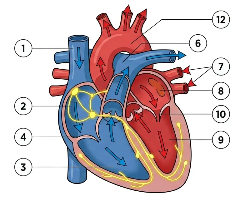
Open image key
Heart Anatomy, Blood Flow, and Electrical Conduction
- 1
- Superior vena cava
- 2
- Right atrium
- 3
- Right ventricle
- 4
- Tricuspid valve
- 6
- Pulmonary artery
- 7
- Pulmonary veins
- 8
- Left atrium
- 9
- Left ventricle
- 10
- Mitral valve
- 12
- Aorta
- Blue arrows
- Deoxygenated blood flowing from the body, through the right heart, to the lungs.
- Red arrows
- Oxygenated blood flowing from the lungs, through the left heart, to the body.
- Yellow paths
- The electrical conduction system (SA Node → AV Node → Purkinje Fibers).
Use the key in this order the first time you study the diagram:
- Trace the blue arrows from the body through the right heart to the lungs.
- Trace the red arrows from the lungs through the left heart to the body.
- Trace the yellow conduction path from the SA node to the AV node and then into the ventricles.
That sequence gives you the structural map and the timing map at the same time.
The Septum Separates the Two Sides
The septum is the muscular wall that separates the right side of the heart from the left side. It matters because it keeps the two circulations distinct, and it also helps you orient the conduction pathway. The AV node sits low in the right atrium near the septal region, and the bundle of His enters the septal area before the signal spreads through the ventricles.
You do not need detailed septal anatomy yet. You do need this picture in your head: the electrical signal starts high in the right atrium, pauses low near the septum, and then moves into the ventricular conduction system.
How Blood Moves Through the Heart
Normal blood flow follows a set route. Oxygen-poor blood returns from the body through the superior vena cava and inferior vena cava, which are the large veins that empty into the right atrium. From there, it moves through the tricuspid valve into the right ventricle.
When the right ventricle contracts, blood moves through the pulmonary valve into the pulmonary arteries and travels to the lungs. In the lungs, carbon dioxide leaves the blood and oxygen enters it. That oxygen-rich blood returns to the heart through the pulmonary veins, which empty into the left atrium.
From the left atrium, blood crosses the mitral valve into the left ventricle. When the left ventricle contracts, it moves through the aortic valve into the aorta and then out to the body.
If you want one clean memory chain, say it in this order:
- Body to right atrium
- Right atrium to right ventricle
- Right ventricle to lungs
- Lungs to left atrium
- Left atrium to left ventricle
- Left ventricle to body
One chamber matters especially for future ECG thinking: the left ventricle has the biggest workload because it pushes blood through the systemic circulation. That larger muscle mass helps explain why ventricular electrical activity usually creates the most prominent part of the tracing.
Valves Keep Blood Moving in One Direction
The heart has four valves that keep flow moving forward. The tricuspid and mitral valves sit between the atria and ventricles, so they are called atrioventricular valves. The pulmonary and aortic valves sit at the ventricular outflow tracts, so they are called semilunar valves.
You do not need valve disease details yet. You do need to know their job. Valves open when pressure pushes blood forward and close to prevent backward flow. That one-way design is what lets the heart act as an efficient pump instead of a sloshing chamber.
The Heart Has To Feed Itself Too
The heart pumps blood to the body, but the heart muscle also needs its own blood supply. The coronary arteries branch off the aorta and deliver oxygen-rich blood to the myocardium, which is the muscular pumping layer of the heart wall.
This matters because cardiac muscle cannot keep working if its own blood supply is reduced. Later, when you learn ischemic changes and conduction problems, you will connect those findings back to the fact that heart tissue needs oxygen like every other tissue.
The Electrical Conduction System Sets the Timing
The ECG exists because the heart is not only a pump. It is also an electrical organ with a built-in conduction pathway. In normal rhythm, the impulse begins in the sinoatrial node, often called the SA node, in the right atrium.
From the SA node, the electrical wave spreads through the atria. That atrial activation is what eventually produces the P wave you will study in the next lessons. The signal then reaches the atrioventricular node, or AV node, near the junction between the atria and ventricles.
After passing through the AV node, the impulse travels into the bundle of His, then down the right bundle branch and left bundle branch, and then through the Purkinje fibers. That organized spread is what allows the ventricles to depolarize in a coordinated way before they contract as a coordinated pump.
The sequence matters more than memorizing every label. In normal conduction, the atria are activated first, there is a short pause at the AV node, and the ventricles activate after that. Later, when you read strips, you will keep asking whether that expected order is still present.
Why the AV Nodal Delay Is Helpful, Not Harmful
The AV node normally slows conduction for a brief moment. This is a feature, not a mistake. That delay gives the atria time to empty blood into the ventricles before the ventricles contract.
If atria and ventricles contracted at the same moment, cardiac filling would be less efficient. The normal sequence is atria first, then ventricles. That timing helps both circulation and ECG interpretation because it creates an expected order you can later recognize on the strip.
Worked Example: Turn the Diagram Into a Strip-Reading Question
Take one normal heartbeat and walk through it in order.
First, the SA node fires high in the right atrium. That activates the atria, so later on a normal strip you should expect atrial activity before ventricular activity. Next, the impulse reaches the AV node and pauses briefly. That gives the ventricles time to finish filling.
Then the signal moves quickly through the His-Purkinje system into both ventricles. Because the ventricles have more muscle mass than the atria, ventricular depolarization usually creates the largest visible deflection on the ECG.
That means this anatomy lesson already gives you a future logic check. If you see ventricular activation with no normal atrial event before it, or if the expected timing between atrial and ventricular activity changes, you already know the beat may not have followed the usual route.
Why This Anatomy Matters When You Read ECGs
This lesson gives you the map behind the tracing. Blood flow tells you which chamber connects to which vessel and why the ventricles have different jobs. The conduction system tells you why atrial activity should come before ventricular activity in normal rhythm.
That makes later ECG questions more logical. If a beat starts in the atria and conducts normally, you should expect an atrial event before a ventricular event. If conduction through the AV node or bundle branches changes, the tracing changes because the pathway changed. If a beat begins low in the heart, the timing and appearance change because the normal route was bypassed.
At a monitoring level, that is the point of this lesson. You are not trying to memorize anatomy for its own sake. You are building the map that lets you say later, “this impulse followed the usual route” or “this impulse started somewhere else or traveled abnormally.”
Don’t Confuse This With
- Right ventricle vs left ventricle - Both are lower pumping chambers, so they are easy to swap early on. The best distinguishing clue: the right ventricle pumps blood to the lungs, while the left ventricle pumps blood to the body.
- Pulmonary arteries vs pulmonary veins - The names feel backward at first. The best distinguishing clue: pulmonary arteries carry oxygen-poor blood away from the heart to the lungs, while pulmonary veins bring oxygen-rich blood back to the heart.
- AV node vs AV valves - Both use the letters A and V, so learners often merge them. The best distinguishing clue: the AV node is electrical tissue that delays conduction, while the AV valves are mechanical structures that direct blood flow.
Lesson Summary
- The heart works as two coordinated pumps, with the right side sending oxygen-poor blood to the lungs and the left side sending oxygen-rich blood to the body.
- Normal blood flow follows a fixed route through the vena cavae, right atrium, right ventricle, lungs, left atrium, left ventricle, and aorta, which gives you the structural map behind bedside monitoring.
- The coronary arteries supply the heart muscle itself, so the myocardium depends on its own blood flow and can develop electrical problems when that supply is impaired.
- Normal electrical activation starts in the SA node, pauses briefly at the AV node, and then travels through the His-Purkinje system to activate the ventricles in an organized way.
- The septum helps you orient both structure and conduction, because the signal pauses near it and then enters the ventricular conduction system from there.
- The most important ECG connection from this lesson is sequence: atrial activation should come before ventricular activation in normal conduction, and later rhythm interpretation depends on recognizing when that sequence changes.
What’s Next
The next lesson shows how depolarization and repolarization become visible waveforms on the ECG. You will take the anatomy and conduction map from this lesson and connect it to the lines and deflections you see on the strip.